Types of color blindness
By Burak. Updated 7 November 2026. About
There are three main groups. Red-green color blindness is by far the most common and has four types: protanopia, protanomaly, deuteranopia and deuteranomaly. Blue-yellow color blindness covers tritanopia and tritanomaly. Complete color blindness, achromatopsia, is very rare. Most color blind people still see many colors; they see fewer differences between them.
How color vision works
The retina has three kinds of cone cells, each tuned to a band of light. L cones respond most to long wavelengths (yellowish red, peaking near 560 nm), M cones to medium wavelengths (green, near 530 nm) and S cones to short wavelengths (blue violet, near 420 nm). The brain compares the three signals: red against green, and blue against yellow.
Color blindness happens when one cone type is missing, or present but tuned to a slightly different wavelength. Then one of those comparisons gets weaker or disappears, and colors that differ only along that line start to look alike.
The types at a glance
| Type | Cone affected | What changes | How common |
|---|---|---|---|
| Deuteranomaly | M (green), shifted | Greens look redder, reds and greens closer | About 5% of men, the most common type |
| Protanomaly | L (red), shifted | Reds look duller and darker | About 1% of men |
| Deuteranopia | M (green), missing | Red, orange, yellow and green fall on one range | About 1% of men |
| Protanopia | L (red), missing | As deuteranopia, and reds look dark | About 1% of men |
| Tritanomaly | S (blue), shifted | Blue and green closer, yellow paler | Very rare |
| Tritanopia | S (blue), missing | Blue and green, yellow and pink mix up | Fewer than 1 in 10,000 |
| Achromatopsia | All cones | No color; light sensitivity, low acuity | About 1 in 30,000 |
What each type sees
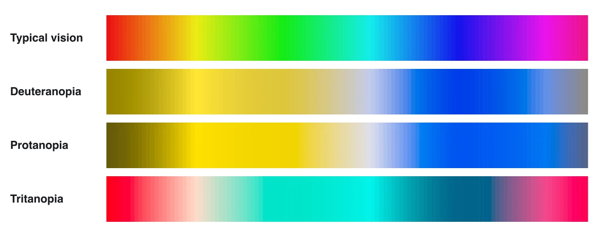
With the two red-green types, the whole red to green half of the spectrum collapses into yellows and browns, while blues stay blue. Protanopia also darkens the red end, which is why a red traffic light or a red laser can look almost black. Tritanopia keeps red and turns the blue to yellow half into cyans and pinks.
Anomaly and anopia
A name ending in -anomaly (protanomaly, deuteranomaly, tritanomaly) means the cone is there but its sensitivity is shifted, so a color difference is smaller than usual. Severity runs from barely noticeable to almost the same as the -anopia form.
A name ending in -anopia (protanopia, deuteranopia, tritanopia) means that cone type is missing, so the difference is gone. The simulator on this site shows the -anopia types at full strength; anomalous vision sits between that and typical vision.
Why men are affected more
The genes for the L and M cone pigments sit next to each other on the X chromosome. Men have one X, so one changed gene is enough. Women have two, so both must carry it. That is why about 1 in 12 men and 1 in 200 women of Northern European descent have red-green color blindness; rates are lower in many other populations.
A woman with one changed gene is a carrier: her vision is usually typical, and each of her sons has a one in two chance of being color blind. The blue cone gene sits on chromosome 7, so tritan types affect men and women equally.
Inherited and acquired
Inherited color blindness is present from birth, affects both eyes the same way and does not change. Color vision can also change later in life, and that kind can affect one eye more than the other and often hits the blue yellow line first.
- Eye disease: glaucoma, diabetic retinopathy, macular degeneration, optic nerve disease.
- Medication: some drugs, such as hydroxychloroquine and ethambutol, need regular color vision checks.
- Age: the lens yellows with age and filters blue light.
- A sudden change in color vision is a reason to see an eye doctor promptly.
How color blindness is tested
Screening usually starts with plate tests such as the Ishihara plates, published in 1917: numbers hidden in dots that only typical color vision separates from the background. Arrangement tests (the Farnsworth D-15) sort colored caps in order. The reference for diagnosis is the anomaloscope, which asks you to match a yellow light with a mix of red and green.
Our online test follows the same ideas with plates generated for screens and a ring test from the Cambridge Colour Test method. A screen is not a calibrated instrument, so it is a screening aid: only an eye care professional can diagnose a color vision deficiency.
An eye doctor explains
An optometrist covers cone types, genetics and acquired color vision loss in under eight minutes.
Questions
What is the most common type of color blindness?
Deuteranomaly, a shifted green cone, affects about 5% of men of Northern European descent. Most people with it can still see red and green, but closer together.
Can women be color blind?
Yes, about 1 in 200 women have red-green color blindness, because they need the changed gene on both X chromosomes. Blue-yellow types affect women and men equally.
Can color blindness be cured?
Inherited color blindness cannot be cured today. Tinted lenses can raise contrast between some colors for some people, and labels, patterns and plain color names help in daily life.
Does color blindness get worse?
Inherited color blindness stays the same for life. A change in color vision later in life can be a sign of eye disease and should be checked by an eye doctor.
Related
- Color blindness simulator
- Color blind test
- What do color blind people see
- Color identifier for color blind people
Sources
- Birch J. (2012), Worldwide prevalence of red-green color deficiency, Journal of the Optical Society of America A 29(3)
- MedlinePlus Genetics, Color vision deficiency and Achromatopsia (US National Library of Medicine)
- National Eye Institute, Types of color vision deficiency
- Machado, Oliveira and Fernandes (2009), A physiologically-based model for simulation of color vision deficiency, IEEE TVCG 15(6)
- Doctor Eye Health (YouTube), How Color Vision Works